26 Sep Clinical Case : Diagnosis and Treatment of Traumatic Bone Cyst with RAYSCAN α
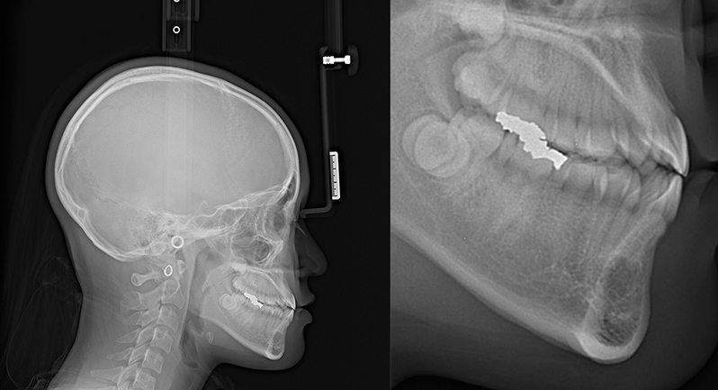
Figure 1. Lateral Cephalometric radiographic image showed a radiolucent lesion in the anterior mandible.
A 15-year-old female presented to our practice for orthodontic treatment to correct a malocclusion associated with mandibular prognathism and an edge-to-edge bite.
At the diagnostic session, we took a lateral cephalometric radiograph (Figure 1) and a panoramic radiograph (Figure 2).
A radiolucent lesion was observed in the anterior mandible, thus a periapical radiograph was obtained (Figure 3). The periapical radiograph demonstrated a radiolucent lesion apical to teeth #s 43,42,41 and 31 (FDI notation).
Download the full text of article.